What is Glaucoma?
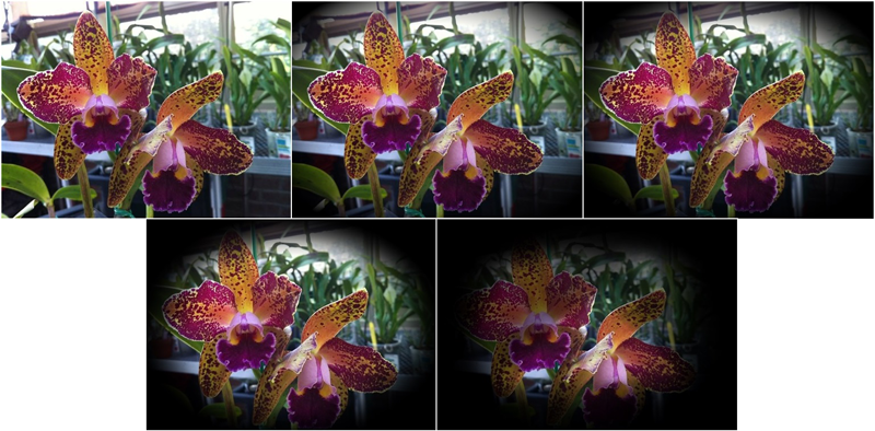
Glaucoma is a condition that causes damage to the optic nerve over time. It is often characterized by elevated intraocular pressure and frequently shows an inheritance pattern. If left untreated, glaucoma can result in irreversible harm to the optic nerve and permanent vision loss. There are several different types of glaucoma:
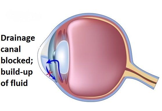
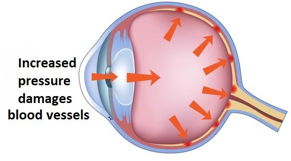
The most common form of glaucoma is Primary Open Angle Glaucoma (POAG) and results from a failure of the eye’s drainage system. This leads to a slow development of increased pressures over time. Initial damage to the optic nerve is typically subtle, having very little effect on vision. Over time, increased damage to the nerve enlarges the visual defects and eventually leads to blindness if left untreated. POAG is considered a medical condition and can be treated with medicated eye drops, laser surgery to stimulate the drainage system, or glaucoma surgery to create a new drainage pathway.
The second most common type of glaucoma, Narrow-Angle or Angle-Closure Glaucoma (NAG), is predominately found in patients who have short eyes. As the lens of the eye ages it thickens to cause crowding in the anterior chamber of the eye and partial blockage of the drainage system. Although the condition develops slowly, eye pressure will build very rapidly if enough of the drainage system is compromised. Common symptoms of an angle closure attack include dramatic and sudden onset of blurred vision, severe eye pain, headaches, nausea or vomiting, and halos. NAG can primarily be treated with a laser to reduce crowding. Alternative treatments can include cataract surgery to reduce crowding or glaucoma surgery to create a new drainage pathway if too much scar tissue is present.
Other types of glaucoma include Pseudoexfolative Glaucoma, Traumatic Glaucoma, Congenital Glaucoma, Normal or Low Tension Glaucoma, and Pigmentary Glaucoma.


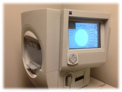
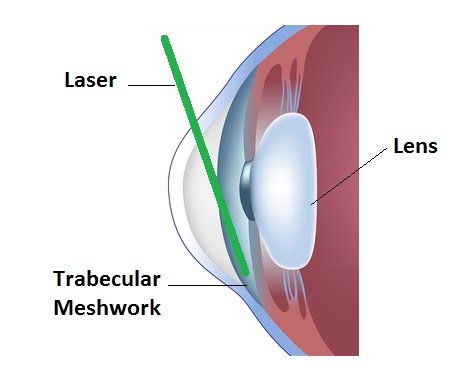 SLT (Selective Laser Trabeculoplasty) is a safe and simple laser treatment that effectively reduces the eye pressure for most patients with Primary Open Angle Glaucoma. The SLT mechanism of effect does not rely on medicines and is considered equal to medicine as a first line therapy for mild to moderate glaucoma. The SLT is performed on a structure known as the trabecular meshwork, which plays a vital role in the balance of internal eye pressure. As a result, the drainage system of the eye is stimulated and function usually improves. Although the effects of the laser will wear off over time, it can be safely repeated. In 2008, Eyes of York was the first facility in York to offer SLT. If you have glaucoma and are using medicated eye drops, ask your doctor about the SLT procedure at Eyes of York.
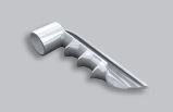 Eyes of York also performs MIGS (Minimally Invasive Glaucoma Surgery) procedures in combination with Cataract Surgery. These devices are implanted during cataract surgery for patients who have mild to moderate open angle glaucoma and are using at least one prescription eye drop to lower their intraocular pressure. These devices work to increase the natural outflow of fluid through the eye by creating a permanent opening in what is known as the trabecular meshwork. They are placed during cataract surgery and cannot be seen, or felt, afterwards. The ultimate goal of these devices is to reduce the pressure in the eye without the need for prescription eye drops. Not all patients with glaucoma are candidates. If you are nearing cataract surgery and have interest in this procedure, ask your doctor if the iStent Inject or Hydrus Microstent are right for you.