What is a medical eye examination?
A medical exam is performed in the same initial manner as a routine eye exam. The reasons for the visit such as symptoms, complaints, and the patient’s diagnoses dictate the level and purpose of the examination and whether the focus will be routine or medical. A patient that presents for a routine eye exam may be interested in new glasses or contacts and may have a diagnosis of myopia with no medical findings. However, the patient appearing for a medical exam may leave with a diagnosis of glaucoma or dry eyes. During a medical eye exam, the structure of the eye may be evaluated by checking for refractive errors, anterior abnormalities, and dilating the pupil. By dilating the pupil the physician is able to take a more detailed look at eye structures past the pupil such as the lens and the retina, evaluating for any possible disease, damage, or irregularities. Medical issues are evaluated more thoroughly with expanded elements as appropriate for the eye exam and possible additional testing for specific conditions.
View Video
How often do I need a medical eye exam?
The frequency of your medical eye exam should be determined by your physician based on your ocular condition and individual treatment plan. Regular follow-ups are crucial when monitoring existing or potentially new conditions concerning the eyes, as some signs and/or symptoms may not be reversible.
Are there diseases and conditions that require routine medical eye exams?
Certain diseases and conditions are more commonly known to require medical eye exams, such as glaucoma, diabetes, and macular degeneration. Other conditions that warrant medical eye exams include cataracts, iritis, hypertension, chronic blepharitis, high myopia, Thyroid disease, Sjogren’s disease, and Lupus. If you have any question regarding what does or does not qualify for a medical exam, contact our office for help.
Are there treatment plans?
At Eyes of York, every patient receives a customized treatment plan based on their individual needs. This plan is formulated after a detailed evaluation of ocular diseases and symptoms as well as overall patient health to determine treatment options and frequency of appointments.
How long will my exam last?
Your exam will likely last 1-2 hours and may include dilation of your pupils.
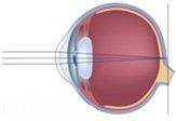
How does the eye work?
The focusing of an image with the human eye is similar to the way a camera focuses a picture. Light rays enter the eye and are bent through both the cornea and lens. The pupil acts like a camera shutter and will open and close to regulate the amount of light that enters the visual system. Most of the bent light rays that enter the eye must come together to a single point on the retina for us to see an image perfectly. If the cornea and lens cannot achieve this together, then glasses or contact lenses are needed. The retina acts like the film in a camera and receives the imprint of the picture, sending it on to the brain for final processing. These several different parts of the eye work together to create the image in front of you. If any of these parts of the eye are not functioning correctly the images you are viewing will be impacted.


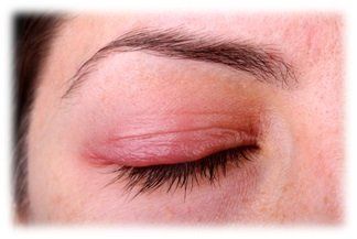 Blepharitis is caused by stagnant meibomian glands and bacteria on the surface of the eyelids and lashes. The bacteria can cause chronic infection or inflammation of the meibomian glands. Symptoms of blepharitis include crusting of the eyelashes when waking, flaking of the skin around the eyes, itchy eyelids, red or swollen eyes, a gritty sensation, and/or tearing.
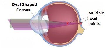
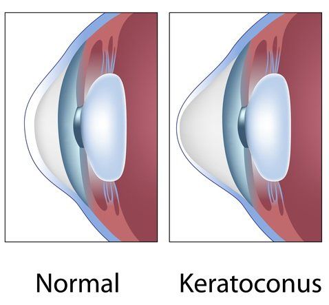 Keratoconus is a condition where the cornea is “cone-shaped” instead of round or dome-like. The irregular cornea will bend light in different directions, creating visual distortion. Keratoconus is a degenerative disorder where symptoms usually become noticeable in the late teens and early twenties. Initial treatments include hard contact lenses which help the cornea maintain a more round shape and bring light into better focus. Some cases of keratoconus progress rapidly for ten to twenty years before ceasing. If the condition progresses, the cornea thins and may require surgery. Common symptoms of keratoconus include blurred vision, light sensitivity, and glare.